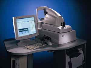
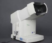
Zeiss ATLAS 9000 Corneal Topography System
- By merging proven ATLAS Placido disk technology with Visante OCT pachymetry, now provides comprehensive anterior and posterior topography
- Proven Placido Disk Technology with patented Cone-of-Focus Alignment System
- SmartCapture Image Analysis Technology analyzes multiple images during alignment and automatically selects the highest quality image
- MasterFit II Contact Lens Software helps streamline the fitting of gas permeable (GP) lenses and guides you through difficult and specialty fits
- Data compatibility with previous generation ATLAS Corneal Topography Systems to facilitate data management and patient follow up
Complete with manuals, software, covers, keyboard, warranty
$
5,475.00
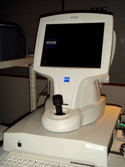
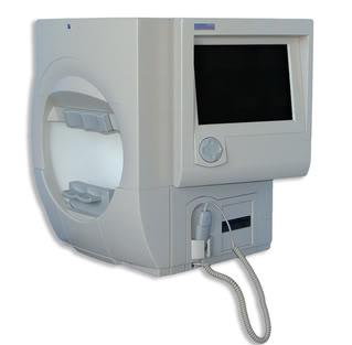
Zeiss Humphrey Atlas 993 Topographer
Humphrey Atlas 993 Corneal Topographer Proven Placido Disk Technology with patented Cone-of-Focus Alignment System, SmartCapture Image Analysis Technology analyzes multiple images during alignment and automatically selects the highest quality image, Integrated, ergonomic design ideally suited for you and your patients, Data compatibility with previous generation ATLAS Corneal Topography Systems to facilitate data management and patient follow up MasterVue Version A10.1 or later
$
3,475.00
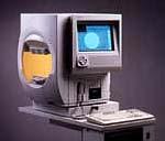
Zeiss GDx VCC Tomographer
- Clinically proven: GDx analysis has been proven to accurately discriminate between healthy and glaucomatous eyes1 and to be predictive of visual field loss2.
- Tracks change over time: The Advanced Serial Analysis feature helps you to evaluate subtle RNFL changes so you can manage glaucoma with more confidence.
- Simple: GDx examinations are easy for your patients and staff. Reports are clear and intuitive.
- Portable: Move your GDx between offices with an optional soft carrying case.
- Fast: Typical exam time ranges from 1 to 3 minutes.
$
4,625.00
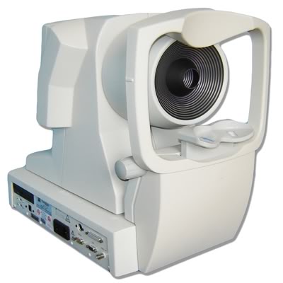
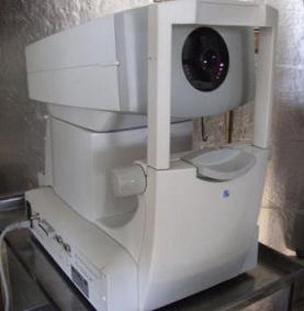
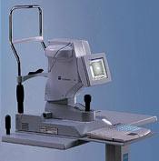
Zeiss Humphrey Atlas 995 Topographer
Description:
The ATLAS Eclipse Model 995 is the most advanced system available.
It offers ultra-low illumination and increased peripheral coverage.
The Model 995 is ideal for high volume corneal and contact lens specialists who require comprehensive and detailed peripheral corneal and pupil assessments.
You can make your ATLAS even stronger with flexible software programs for corneal analysis, refractive surgery evaluation and contact lens fitting.
$
3,990.00
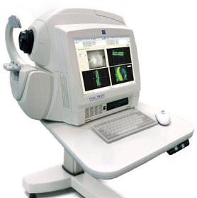
ZEISS Stratus OCT-3 OCT 3 Ophthalmology General
Description:
The Stratus OCT incorporates optical coherence tomography technology to provide comprehensive imaging and measurement of glaucoma and retinal disease. Stratus OCT is the gold standard in vivo imaging device for the posterior segment and offers proven reproducibility for disease management.
The Stratus OCT provides real-time cross-sectional images and quantitative analysis of retinal features to optimize the diagnosis and monitoring of retinal disease and for enhanced pre- and post-therapy assessment. The device is beneficial for evaluation of cataract patients, pre- and post-operatively, and for the assessment of early signs of glaucoma and glaucomatous change.
$
13,090.00
Humphrey Zeiss 750i Perimeter Visual Field
- The Gold Standard in automated perimetry for enhanced clinical confidence
- HFA-NET Pro software provides connectivity to electronic medical records (EMR) systems or your office network
- Guided Progression Analysis(GPA) identifies statistically significant changes in visual field threshold sensitivity automatically
- SITA SWAP software reduces blue-yellow threshold test time to just 3 to 6 minutes, providing a clinically practical tool for early glaucoma detection
- Advanced analysis tools backed by decades of research and clinical validation
$
6,190.00
Humphrey Zeiss 740i Perimeter Visual Field
Includes: Printer and Wheel Chair accesible power Table
$
5,475.00
Humphrey HARK-599 Autorefractor
Description:
The HARK 599 is the first full-featured combination automatic refractor/keratometer that enables both subjective and objective refraction with low-contrast, glare and visual acuity testing. The HARK allows for immediate patient feedback on the prescription.
Automatic alignment and tracking allow the instrument to follow shifts in eye movement during testing, while speeding up routine examination time, improving patient comfort level and achieving accurate refractions. Automatic keratometry provides precise keratometric readings, including Delta Ks.
The operator control panel can be positioned at either 90° or 180° from the patient to meet any office space requirements while encouraging communication between patient and operator.
$
2,640.00
Humphrey FDT Visual Field
Description:
The Humphrey FDT Perimeter uses Welch Allyn Frequency Doubling Technology to provide a clinically verified, fast and affordable means of detecting early visual field loss. The perimeter is the ideal glaucoma screening device because it conducts supra-threshold testing in only 35 seconds per eye.
Remarkably affordable, the FDT Perimeter brings glaucoma testing to any size practice, even the smallest. In addition to fast supra-threshold testing it also conducts full threshold testing complete with statistical analysis in about 4 minutes.
$
2,825.00
Zeiss IOL Master
Description:
Norwich Ophthalmology Group uses the Zeiss IOLMaster, the first device that quickly and precisely measures boundaries for intraocular lens (IOL) implantation without ever touching the cornea.
If a patient’s natural lens of the eye is removed, due to cataracts or vision correction purposes, an IOL is then implanted into the eye. This IOL’s “power” or strength needed to give the patient good vision, has been very carefully calculated by the surgeons at Norwich Ophthalmology Group. Though the instrument previously used to take these measurements was accurate and dependable, the IOLMaster has highly refined this measurement.
The IOLMaster is yet another technological advancement that Norwich Ophthalmology Group surgeons utilize to ensure high accuracy, which in turn equals superior post-operative visual outcomes.
$
5,925.00
Zeiss Cirrus 4000 OCT HD
Technical data
OCT Scanning - Axial resolution: 5 μm (in tissue)
- Transverse resolution: 15 μm (in tissue)
- Scan speed: 27,000 A-scans per second
- A-scan depth: 2.0 mm (in tissue), 1024 points
- Optical source: superluminescent diode (SLD), 840 nm
Fundus Imaging - Line scanning ophthalmoscope (LSO)
- Live during scanning
- Transverse resolution: 25 μm (in tissue)
- Optical source: superluminescent diode (SLD), 750 nm
- Field of view: 36° x 30°
Scan Patterns - Macular Cube 200 x 200 Combo: 200 horizontal scan lines comprised
of 200 A-scans
- Macular Cube 512 x 128 Combo: 128 horizontal scan lines comprised
of 512 A-scans
- 5 Line Raster: 4096 A-scans per B-Scan (adjustable length, spacing and
orientation)
Focus Adjustment Range - –20D to +20D (diopters)
Fixation - Internal and external
Computer - Windows® XP Pro
- High-performance multi-core processor
- Internal storage: > 80,000 scans
- CD-RW, DVD-ROM drive
- Integrated 15” color flat panel display
Pupil Size Requirement - ≤ 2.0 mm (≥ 3.0 mm optimal for LSO)
Dimensions/Weight
(Instrument Only)
- 25.6 L x 17.3 W x 20.9 H (in); 65 L x 44 W x 53 H (cm)
- 83 lbs; 37.6 kg
Electrical 100–120V~, 50/60Hz, 5A
220–240V~, 50/60Hz, 2.5A
$
16,175.00
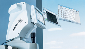
Carl Zeiss IOLMaster 500
Feature:
- Increased Speed: In individual practice studies, the new IOLMaster 500 has been shown to complete all measurements in as few as 80 seconds*.
- Dual-mode Measurements: Axial length and keratometry calculations can be collected simultaneously, at the push of a button.
- Composite Signal Filtering: New sound filtering technology excludes poor readings and increases the percentage of cataract patients that can be measured.
- Seamless Data Integration with the Synergy Ultrasound A-scan: This time-saving feature allows easy workflow integration of A-scan ultrasound biometry for difficult cases, such as extremely dense cataracts.
- Easy Data Transfer to the Holladay Consultant Program: The Holladay II formula has become very popular among physicians in North America.
- New Graphical User Interface: With green, red and yellow "traffic light" reporting, technicians of all experience levels can achieve highly accurate diagnostic results.
$
7,950.00
